From Spines to Skulls: Unveiling the Secrets of the Skeletal System
Introduction
The human skeletal system is an intricate framework that provides structure, protection, and support to the body. Comprising 206 bones in the average adult, it serves as the foundation for more complex biological systems. The skeletal system is not merely a collection of bones; it is a dynamic organ system that plays roles in movement, mineral storage, and blood cell production. In this article, we will explore the various functions of the skeletal system, examine its anatomical structure from vertebrae to cranium, and uncover the evolutionary adaptations that have allowed it to thrive.
Anatomy of the Skeletal System
1. Bone Types
There are four main types of bones in the human body:
- Long Bones: Primarily found in the limbs, long bones like the femur and humerus are longer than they are wide and are crucial for mobility.
- Short Bones: These are approximately as wide as they are long, allowing for stability and support. Examples include carpals and tarsals.
- Flat Bones: Usually protecting vital organs, flat bones such as the skull, ribs, and sternum provide surface area for muscle attachment.
- Irregular Bones: These bones have complex shapes that do not fit into the other categories, such as the vertebrae and pelvic bones.
2. Major Bone Structures
The skeletal system can be divided into two main parts: the axial skeleton and the appendicular skeleton.
Axial Skeleton
-
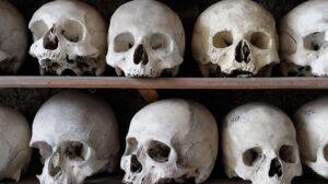
Skull: Composed of 22 bones, the skull protects the brain and supports facial structures. It consists of the cranium and facial bones.
-
Vertebral Column: The spine consists of 33 vertebrae, divided into cervical, thoracic, lumbar, sacral, and coccygeal regions. Each part has distinct functions, with the cervical region allowing for head movement and the lumbar region enduring weight-bearing activities.
- Ribs and Sternum: Protecting the thoracic cavity, the rib cage consists of 12 pairs of ribs attached to the breastbone (sternum) at the front.
Appendicular Skeleton
-
Upper Limbs: Including the humerus, radius, ulna, carpals, metacarpals, and digits, the upper limbs allow for a wide range of motion.
-
Lower Limbs: The femur, tibia, fibula, tarsals, metatarsals, and phalanges form the lower limbs, which are crucial for locomotion.
- Pelvic Girdle: Comprising the hip bones, this structure connects the spinal column to the lower limbs and supports the weight of the upper body while standing and walking.
Functions of the Skeletal System
1. Support
The skeleton provides structural support for the body, allowing it to maintain its shape. Without the skeleton, muscles and organs would collapse under the force of gravity.
2. Protection
Bones safeguard vital organs from injury. For instance, the skull encases the brain, while the rib cage shields the heart and lungs.
3. Movement
Joints formed by bones interacting allow for movement. Muscles attach to bones via tendons, and when muscles contract, they pull on bones to create movement at the joints.
4. Mineral Storage
Bones act as a reservoir for minerals, particularly calcium and phosphorus. The body can withdraw these minerals from bones to maintain vital metabolic functions.
5. Blood Cell Production
Hematopoiesis, or the production of blood cells, occurs in the bone marrow found within certain bones (e.g., the femur, humerus, and vertebrae). This process is vital for maintaining adequate blood supply and immune function.
6. Energy Storage
Bone tissue also contains adipocytes, which store lipids. This stored fat serves as an energy reserve that the body can utilize when required.
Development and Growth of the Skeletal System
1. Embryonic Development
The skeletal system begins forming during the embryonic stage, originating primarily from mesodermal cells. Initially, the skeleton is cartilaginous and gradually ossifies through endochondral and intramembranous ossification processes.
2. Growth Plates
Longitudinal growth of bones occurs at the epiphyseal plates or growth plates located at the ends of long bones. These plates allow for lengthening until puberty when they fuse, ceasing further growth.
3. Remodeling
The skeleton is continually remodeled throughout life, a process that involves the resorption of old bone and the formation of new bone. This adaptive remodeling is influenced by mechanical stress, hormonal levels, and dietary factors.
Common Skeletal Disorders
1. Osteoporosis
A condition marked by reduced bone density, osteoporosis increases the risk of fractures. It is often related to aging, hormonal changes, and nutritional deficiencies.
2. Arthritis
Inflammation of the joints can lead to various forms of arthritis, including osteoarthritis and rheumatoid arthritis, which significantly impact mobility and quality of life.
3. Scoliosis
This condition is characterized by an abnormal curvature of the spine, which can result from congenital factors, neuromuscular conditions, or idiopathic causes.
4. Fractures
Fractures can occur due to trauma, overuse, or underlying bone diseases. Common types include simple, compound, and stress fractures, each requiring different management approaches.
The Evolution of the Skeletal System
The skeletal system is a product of millions of years of evolution, adapting to various environmental pressures. From the early vertebrates to the diverse forms seen in modern mammals, the skeletal structure has evolved in accordance with locomotion, feeding, and reproductive needs.
1. Primitive Skeletal Forms
The earliest vertebrates possessed a cartilaginous skeleton, which provided greater flexibility in aquatic environments. As organisms adapted to life on land, skeletons gradually evolved to become more robust and supportive.
2. The Rise of Mammals
Mammalian skeletons exhibit adaptations that support endothermy and increased mobility. For instance, limb bones have become elongated and more specialized, allowing for diverse locomotion.
Conclusion: The Living Skeleton
The skeletal system is not a static structure but a living organ, constantly adapting to the needs of the body. As research into bone biology advances, understanding the complexities of skeletal health and disease becomes increasingly important. This knowledge will undoubtedly lead to better treatments and preventive strategies for maintaining skeletal integrity throughout life.
In understanding the journey from spines to skulls, we unveil not only the anatomy of the skeletal system but also its intricate, multifaceted roles in human health and vitality.
References
[1] "Human Anatomy & Physiology" — Marieb, E. N., & Hoehn, K. [2] "Orthopedics: Principles and Practice" — Rothman, R. H., & Simeone, F. A. [3] "Skeletal System: Structure and Function" — Getz, J. [4] "Bone Density and Osteoporosis: A Comprehensive Review" — Schuit, S. C., et al. [5] "The Role of the Skeleton in Homeostasis" — Canalis, E., et al. [6] "Evolutionary Biology of the Skeletal System" — Hall, B. G. [7] "Joint Health and Disease Management" — Thomas, A., & Evans, R. [8] "Fractures: Diagnosis and Treatment" — Nordin, B. E. C., & Frankel, V. H.By exploring each aspect of the skeletal system, from developmental biology to its evolutionary history, we gain a deeper appreciation of this remarkable aspect of human anatomy, providing insights not just into our physical structure but the very essence of our biological heritage.













Add Comment