


Unlocking the Body’s Framework: Understanding the Skeletal System Through Diagrams
Introduction
The human skeletal system is a marvel of biological engineering. It provides the body with structure, protects vital organs, and allows for movement—functions essential for survival and quality of life. Understanding this intricate framework is fundamental not only for professionals in health and science but also for anyone interested in human anatomy. This article explores the skeletal system using diagrams to visualize its components, relationships, and functions.
1. Overview of the Skeletal System
The human skeletal system comprises 206 bones in an average adult, with this number varying in children due to the presence of growth plates. The skeleton can be broadly categorized into two parts: the axial skeleton and the appendicular skeleton.
1.1 The Axial Skeleton
The axial skeleton includes the skull, vertebral column, and rib cage. It serves as the central framework that supports the body.
- Skull: Protects the brain and forms the face.
- Vertebral Column: Supports the head and torso, providing flexibility and protection to the spinal cord.
- Rib Cage: Protects the heart and lungs while aiding in respiration.
1.2 The Appendicular Skeleton
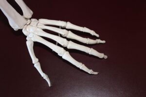
The appendicular skeleton consists of the bones of the limbs and girdles. This segment facilitates movement and interaction with the environment.
- Upper Limbs: Comprise the humerus, radius, and ulna; allow for a wide range of motion.
- Lower Limbs: Include the femur, tibia, and fibula; support body weight and aid in locomotion.
- Pelvic Girdle: Connects the lower limbs to the axial skeleton, providing stability.
Diagram 1: Major Components of the Human Skeleton
Summary: A labeled diagram outlining the axial and appendicular skeleton will give readers a visual context for the discussion.
2. Bone Structure and Classification
Bones can be classified into several types based on their shape and function. Understanding these types is vital for comprehending how the skeletal system operates.
2.1 Types of Bones
- Long Bones: e.g., femur and humerus; characterized by a long shaft and are primarily involved in movement.
- Short Bones: e.g., carpals and tarsals; provide stability while allowing for some movement.
- Flat Bones: e.g., skull bones and sternum; protect internal organs and provide surfaces for muscle attachment.
- Irregular Bones: e.g., vertebrae; have complex shapes that serve specific purposes.
2.2 Bone Structure
Each bone consists of several layers:
- Compact Bone: Dense outer layer that provides strength.
- Spongy Bone: Lighter, porous inner layer that houses bone marrow.
- Periosteum: A protective membrane covering the outer surface of the bone, containing blood vessels and nerves.
Diagram 2: Structure of a Long Bone
Summary: This visual will highlight the different layers and components of a long bone, aiding in the understanding of its anatomy.
3. Joint Types and Functions
Joints, where two or more bones meet, play a crucial role in the skeletal system by facilitating movement. The type of joint determines the range and type of motion possible.
3.1 Types of Joints
- Fibrous Joints: Immovable joints held together by dense connective tissue (e.g., sutures in the skull).
- Cartilaginous Joints: Allow limited movement (e.g., intervertebral discs).
- Synovial Joints: Freely movable joints characterized by a fluid-filled cavity (e.g., hip and knee joints).
3.2 Synovial Joint Examples
- Hinge Joints: Permit back-and-forth motion (e.g., elbow).
- Ball-and-Socket Joints: Allow rotational movement (e.g., shoulder).
- Pivot Joints: Enable rotation (e.g., cervical vertebrae).
Diagram 3: Types of Joints
Summary: An illustrative chart categorizing different joint types will help clarify their functions and locations.
4. The Role of the Skeletal System
Understanding the functions of the skeletal system is key to appreciating its importance to overall health.
4.1 Support
The skeleton provides a rigid framework that supports the body’s shape and facilitates the attachment of soft tissues and organs.
4.2 Protection
Vital organs are shielded by bone. For example, the skull protects the brain, while the rib cage guards the heart and lungs.
4.3 Movement
Bones act as levers when muscles contract, allowing for various motions. Joints enable flexible movement, which is critical for daily activities.
Diagram 4: Functions of the Skeletal System
Summary: A diagram summarizing the functions of the skeletal system will reinforce the information presented.
5. Bone Health and Maintenance
Maintaining bone health is crucial for longevity and quality of life. Various factors influence bone development and maintenance.
5.1 Nutrition
- Calcium: Essential for bone strength; found in dairy, leafy greens, and fortified foods.
- Vitamin D: Facilitates calcium absorption; obtained from sunlight and dietary sources.
- Protein: Important for bone structure; sources include meats, beans, and nuts.
5.2 Exercise
Weight-bearing activities stimulate bone growth and density. Exercises such as walking, running, and strength training are beneficial.
5.3 Lifestyle Factors
- Smoking and Alcohol: Both negatively impact bone health.
- Hormonal Changes: Particularly in women, can lead to conditions like osteoporosis.
Diagram 5: Factors Influencing Bone Health
Summary: A graphic showcasing the various lifestyle factors affecting bone health can be an effective learning tool.
6. Common Skeletal Disorders
Understanding disorders affecting the skeletal system can provide critical insights into the importance of bone health.
6.1 Osteoporosis
A condition characterized by weakened bones, increasing the risk of fractures. It often goes unnoticed until a fracture occurs.
6.2 Arthritis
Inflammation of the joints leading to pain, swelling, and decreased mobility. Different types include osteoarthritis and rheumatoid arthritis.
6.3 Scoliosis
A condition causing abnormal curvature of the spine, which can affect posture and may require treatment.
Diagram 6: Common Skeletal Disorders
Summary: Illustrative diagrams showcasing the anatomical changes in these conditions help convey their implications effectively.
7. Conclusion
Unlocking the mysteries of the skeletal system offers profound insights into how our bodies function. Using diagrams to visualize this complex framework aids in understanding its anatomical structure and functions. By promoting awareness of bone health and common disorders, we empower individuals to take proactive steps in maintaining their skeletal system, thereby enhancing their overall quality of life.
This article provides a foundation for further exploration into the skeletal system, encouraging curiosity and learning through visual aids and diagrams. Understanding our body’s framework is not just an academic pursuit; it’s a vital part of fostering a healthier lifestyle.
References
- Marieb, E. N., & Hoehn, K. (2018). Human Anatomy & Physiology. Pearson.
- Tortora, G. J., & Derrickson, B. H. (2017). Principles of Anatomy and Physiology. Wiley.
- Hall, B. K., & Haff, G. G. (2021). Bone Biology and Bone Disorders. Springer.
- Melton, L. J., Khosla, S., Crowson, C. S., O’Fallon, W. M., & Riggs, B. L. (2000). "The prevalence of osteoporosis and fractures in older women: a population-based study." The Journal of Bone and Mineral Research, 15(12), 2267-2277.
- Lane, J. M., & Finkelstein, J. W. (2019). "Osteoporosis: Dual-energy x-ray absorptiometry". Bone Health and Osteoporosis: A Guide for Clinicians. 1-37.
This article serves as an introduction to understanding the skeletal system through visual tools, aiming to educate and encourage further inquiry into the human body’s remarkable architecture.


Add Comment