


The Backbone of Anatomy: Exploring the Skeletal System
Introduction
The human skeletal system is a marvel of biological engineering, providing not just a framework for the human body, but also facilitating movement, protecting vital organs, and producing blood cells. Comprised of 206 bones in adults, the skeletal system is a complex structure that interacts with other bodily systems to maintain overall health and functionality. This article aims to delve into the intricacies of the skeletal system, supported by detailed diagrams and modern insights into its anatomy and physiology.
Overview of the Skeletal System
Composition of the Skeletal System
The skeletal system is primarily composed of bones, cartilage, ligaments, and joints.
-
Bones: Composed of a dense connective tissue that contains minerals (such as calcium and phosphorus), bones are categorized into two types:
- Compact bone: The dense, outer layer that provides strength.
- Spongy bone: A lighter, porous tissue found inside bones that aids in weight reduction and shock absorption.
-
Cartilage: A flexible tissue that reduces friction and absorbs shock at joints. It also provides structure in other areas, like the nasal septum and the ear.
-
Ligaments: Tough bands of connective tissue that connect bones to other bones, helping to stabilize joints.
-
Joints: The connections between bones that allow for movement. Categories include:
- Synovial: Freely movable (e.g., knees, elbows).
- Cartilaginous: Limited movement (e.g., spine).
- Fibrous: Immovable (e.g., skull sutures).
Functions of the Skeletal System
The skeletal system serves several critical functions:
- Support: Provides a structural framework for the body, holding organs in place.
- Protection: Shields vital organs (e.g., the skull protects the brain, the rib cage protects the heart and lungs).
- Movement: Serves as the attachment points for muscles, facilitating mobility.
- Mineral Storage: Stores minerals like calcium and phosphate, which are critical for numerous bodily functions.
- Blood Cell Production: Houses bone marrow, which produces red blood cells, white blood cells, and platelets.
Detailed Components of the Skeletal System
Axial Skeleton
The axial skeleton consists of 80 bones and is divided into three major regions:
-
Skull: Comprised of 22 bones (including the cranial and facial bones) that encase the brain and form the structure of the face.
-
Vertebral Column: Made up of 33 vertebrae (including cervical, thoracic, lumbar, sacral, and coccygeal vertebrae) that protect the spinal cord and provide posture.
-
Thoracic Cage: Consists of the sternum and ribs, protecting the heart and lungs while assisting in respiratory mechanics.
Diagram 1: (Axial Skeleton Diagram)
Appendicular Skeleton
The appendicular skeleton includes 126 bones and is responsible for movement and interaction with the environment. It is further divided into:
-
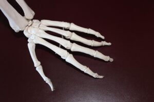
Upper Limbs: Comprising the humerus, radius, and ulna, along with carpals, metacarpals, and phalanges which facilitate arm and hand movement.
-
Lower Limbs: Comprising the femur, tibia, and fibula, along with tarsals, metatarsals, and phalanges that enable walking and running.
-
Pelvic Girdle: Composed of the hip bones (ilium, ischium, and pubis), which connects the lower limbs to the axial skeleton and supports bodily weight.
Diagram 2: (Appendicular Skeleton Diagram)
The Development of the Skeletal System
The development of the skeletal system occurs over time and follows distinct stages:
Embryonic Development
The skeleton starts to develop during the third to fourth week of gestation, initially forming from cartilage templates that gradually ossify (turn into bone) through endochondral ossification.
Childhood to Adolescence
During growth, bones undergo continual remodeling in response to mechanical stress. Factors such as nutrition and physical activity can impact bone growth, density, and strength.
Aging and Bone Health
As individuals age, bone density typically decreases, which can lead to conditions like osteoporosis. It’s critical to maintain bone health through diet (rich in calcium and vitamin D) and exercise.
Common Skeletal Disorders
Maintaining healthy bones is essential to avoid various disorders:
-
Osteoporosis: A condition where bones become weak and brittle, often due to aging or deficiency in calcium.
-
Arthritis: Inflammation of the joints causing pain and stiffness. Common forms include osteoarthritis and rheumatoid arthritis.
-
Fractures: Breaks in bones due to trauma or disease, requiring proper care for healing.
-
Scoliosis: A lateral curvature of the spine that can occur during growth, requiring monitoring and sometimes bracing or surgery.
Modern Techniques for Studying the Skeletal System
Imaging Techniques
-
X-ray: A primary tool for visualizing bone structure, identifying fractures, and assessing joint health.
-
MRI (Magnetic Resonance Imaging): Useful for examining soft tissues, cartilage, and bone marrow.
-
CT Scans (Computed Tomography): Provides detailed cross-sectional images that help in the diagnosis of skeletal disorders.
Advanced Research
-
Bone Density Scans: Utilized to assess the risk of fractures and monitor conditions like osteoporosis.
-
Genetic Studies: Investigating hereditary conditions influencing bone growth and health.
Conclusion
The skeletal system is not merely a set of bones; it is a dynamic and indispensable system that plays a fundamental role in overall human health and functionality. Understanding its components, functions, and common disorders equips individuals with the knowledge necessary for maintaining a healthy lifestyle.
References
- “Human Anatomy & Physiology.” 11th Ed. Pearson, 2018.
- “Principles of Anatomy and Physiology.” Tortora, G. J., & Derrickson, B. 15th Ed. Wiley, 2021.
- “Orthopedic Secrets.” Kelikian, A. S. 5th Ed. Elsevier, 2017.
- “Fundamentals of Anatomy & Physiology.” 11th Ed. F. A. Davis Company, 2019.
- Modern Imaging Techniques in Musculoskeletal Disorders. Journal of Bone and Joint Surgery, Volume 99, Issue 7, 2017.
This article provides a comprehensive overview of the skeletal system, featuring insightful diagrams and drawing from both classical knowledge and modern advancements in medical science. The dynamic nature of the skeletal system emphasizes its importance and necessitates continued research and education to foster better health outcomes for individuals.


Add Comment